Basic Bacterial Morphology
Bacteria and fungus (mold and yeast) grow in a variety of shapes, sizes and colors. Although it is difficult to make a definite identification of cultures growing in broth or solid media many have distinguishing characteristics that give us clues to identification. Many Microbiologist can make a tentative identifications by growth characteristics but definitive id requires further testing (ie Gram stain, chemical testing, etc).
 |
| The palm of a hand print print done on TSA agar. There is a mix of different textured and colored colonies on the plate some bacterial some yeast. By looking at color, texture, growth times and various growth temperatures you can get an idea as to what is on the plate. Ex: the orange colonies are yeast and tend to grow better at room temperature or refrigerated temperatures where the large colony on the left (Bacillus spp) and the white colonies (Staphylococcus) grows well at body temperature. Further testing needs to be done, starting with Gram staining, for a positive ID. |
Isolation of Colonies: Streak Plate
The first step in identifying an organism is to isolate the individual bacteria/yeast/fungus. This is done by doing a streak plate for isolation.
The first step in identifying an organism is to isolate the individual bacteria/yeast/fungus. This is done by doing a streak plate for isolation.
 |
| Inoculating loop with uninocualted Trypticase Soy Agar (TSA) |
The basis of the streak plate is to dilute the sample on the agar plate by dividing the plate into either 3 or 4 quadrents with the initial streak being at full strength. After the initial streak the loop is flamed before each streak which dilutes the sample down to individual colonies.


 |
Example of a streak plate with no isolation. Serratia marcessens was streaked onto TSA but after 24 hrs of growth there was no isolation indicating poor technique. More then likely the loop was not flamed between streaks so there was no dilution of the specimen.

Examples of a 3 quadrant streak plate done to isolate colonies. The goal is to get isolated colonies, one bacteria = one colony. So each individual colony can be subcultured onto another plate and that can be considered a pure colony.
Isolation of Colonies from a Mixed Culture: Streak Plate |
| Streak plate with isolated colonies of a mixed culture of Micrococcus luteus (yellow, circular colonies) and E. coli (white colonies). |
 |
Streak plate of a mixed culture Serratia marcescens (red) and Staphylococcus aureus (whites)grown on TSA for 48 hrs. at 37 degree's C then held at room temp for 24 hrs. |
Growth Characteristics in different Media
After the organism is isolated there are a number of different ways of inoculating media to look at different characteristics.
Broth Tubes:
Growth in broth tubes can be characterized in the following way:
-Uniform fine turbidity: Finely dispersed growth throughout
- Pellice: Thick, padlike growth on the surface. Many times the organism will sink or fall after 48 hrs growth forming what might look like a pellet or sediment at the bottom of the tube
- Sediment: Concentration of growth at the bottom of the broth tube
-Flocculant: Flaky aggregates dispersed thought out (not pictured)

Broth media can be used to look at how an organism grows in liquid. Far left Staphylococcus aureus: grows confluently throughout the tube (turbid). Center Mycobacterium smegmatis: grows only at the tops and forms a pellicle. Far right Bacillus subtilis: grows then settles out forming a pellet or sediment.

Shape and Texture
Many bacterial, fungal, yeast colonies all have distinctive growth patterns on media and can be tentatively identified by what they look like on an agar plate. Many organisms have distinctive growth patterns make it easier to make a tentative identification.
Form:
-Filiform: Continuous, threadlike growth with smooth
edges
-Echninulate: Contunuous, threadlike growth with irregular
edges
-Beaded: Nonconfluent to semiconfluent colonies
-Effuse:
Thin, spreading growth
-Arborescent:
Treelike growth
-Rhizoid: Rootlike growth
 |
| Unknown contaminant on agar with rhizoid, circular, raised growth with exeduate or droplets being produced. Although this looks like a mould it is more then likely a Bacteria known as Streptomyces. |
 |
| Another unknown contaminant that looks like a mould but is a Streptomyoces. Spreading lobate irregular edges with a grainy like appearance. |

Above is a single colony of Bacillus mycodies growing on a TSA plate. The margins are swirling growth patterns, always curls to the left and can be considered Arborecent or Rhizoid with spreading growth. One colony covered an entire TSA plate.

Close up of Bacillus mycodies branching growth.

Unknown organism that has Rhizoid or Arborecent form.
 Unknown organism, most likely a yeast or bacillus, with a beaded, raised, yellow appearance with lobate/irregular edges.

Above is a single colony of Bacillus mycodies growing on a TSA plate. The margins are swirling growth patterns, always curls to the left and can be considered Arborecent or Rhizoid with spreading growth. One colony covered an entire TSA plate.

Close up of Bacillus mycodies branching growth.


 |
| Unknown yeast from and environmental swab, more then likely Rhodotrula spp, highly mucoid with smooth edged with few undefined circular raised colonies. |

Left and bottom: Straight line inoculation of Mycrobacterium smegmatis after a weeks growth at 37 degree's C. The growths looks dry (matt) and cracked and is known as friable.


Right: Straight line inoculation of Micrococcus luteus. Growth is mucoid, yellow (pigment) and is echinulate (irregular edges with continuous growth)


On the Right: Straight line inoculation of Staph aureus with filifrom growth
On the left: Straight line inoculation of Bacillus subtilis with effuse (spreading) growth.

Left: Straight line inoculation of Serratia marcesens with filiform growth.

Unknown organism with lobate/irregular edges, raised center and yellow pigment.

Unknown yeast contaminant with entire well defined edges, raised center, pigmented and mucoid (shiny)
Plates:
1). Size:
-Pinpoint
-small
-moderate
-large
2). Pigment: color of colony.
3). Form:
-Circular: Unbroken peripheral edge
-Irregular: Indented perpherial edge
-Rhizoid: Rootlike perpherial edge
4). Margin:
-Entire:
Sharply defined, even
-Lobate: Marked indentations
-Undulate: Wavy indentations
-Serrate: Toothlike appearance
-Filamentous: Threadlike, spreading edge
5).
Elevation:
-Flat: Elevation not discernable
-Raised: Slightly elevated
-Convex: Dome-shaped elevation
-Umbonate: Raised, with
elevated convex central region
Right: Straight line inoculation of Micrococcus luteus. Growth is mucoid, yellow (pigment) and is echinulate (irregular edges with continuous growth)


On the Right: Straight line inoculation of Staph aureus with filifrom growth
On the left: Straight line inoculation of Bacillus subtilis with effuse (spreading) growth.

Left: Straight line inoculation of Serratia marcesens with filiform growth.
 |
| Unknown organism with lobate/irregular edges, raised center and yellow pigment. |
 |
| Unknown yeast contaminant with entire well defined edges, raised center, pigmented and mucoid (shiny) |
Plates:
Plates
After 24 hours with the exception of
M. luteus and M. smegmatis
Margins Elevation
Form Pigment
Size
E. coli undulate raised
irregular white/opaque
moderate
B sub/cereus lobate
raised irregular/rhizoid white/dull/dry large/spreading
S. marcesens entire
umbonate circular red/mucoid moderate
center raised area
M. smegmatis lobate umbonate irregular crusty/dry small
yellow
M. luteus entire
convex circular yellow/mucoid pinpoint
small
S. aureus entire
raised circular white(-)/yellow(+) moderate
 |
| Streak plate of mixed culture E.coli (undulate, white/opaque, rasied) and Micrococcus luteus (entire, circular raised, yellow glossy or mucoid) |
 |
| Streak plate of Bacillus subtilis (labate, raised,dry/matt, irregular) |
 |
| Streak plate of mixed culture Staphylococcus aureus (white, entire, circular and opaque) and Serratia marcessens (enitire, circular, red pigment) |
 |
| Streak plate of M. Smegmatis after 72 hrs growth with lobate edges, umbonate center (pinpoint raised area in the center), crusty/dry and yellow. |
Pigmented Bacteria: Chromagenic bacteria that produce color
When looking at colonies optical characteristics such as color and transparency are also noted.
Optical
Characteristics:
-Opaque: no
light transmission
-translucent:
partial transmission
-Transparent:
full transmission


Streak plate of a mixed culture Serratia marcescens grown on TSA for 48 hrs. at 37 degree's C then held at room temp for 24 hrs. Serratia marcescens is circular, mucoid and has a red to orange pigment due to the production of prodigiosin. Usually the red coloration dose best at reduced temperatures so after initial grown at 37 degree I will hold the plate at room temperature for 24 hrs to develop the intense red coloration or grow a pure culture at room temp for 24 hrs to develop the intense red color. Staphylococcus is the white circular colonies on the plate.
 |
Golden yellow/orange (carotenoid pigment), mucoid, circular colonies of Sarcinia aurantiaca grown at room temperature. These are Gram (+) coccus bacteria that grow in cubes of 8. |
 |
Yellow (carotenoid pigment), mucoid, circular colonies of Micrococcus luteus. Gram (+) coccus. |
 |
Purple (violacein, also is a naturally occuring antibiotic), mucoid (shiny) circular colonies of Chromobacterium violaceum grown at room temperature (25 degree's C) for several day's. This is a Gram (-) coccobacillus. |
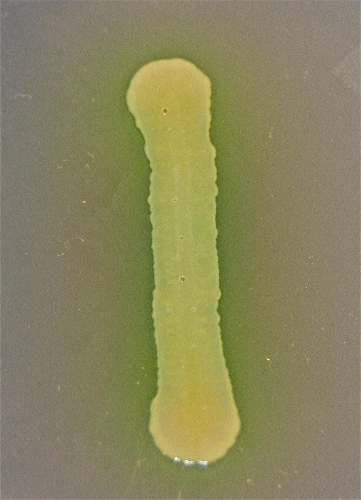 |
| Some organism produce a pigment that spreads out into the surrounding media known as a diffusible pigment. Above is a straight line inoculation of Pseudomonas aeroginosa which produces a green diffusible pigment that can be around the growing colony. |
All images are copy righted


Hi, I am just wondering why do you have to plate the new microbes over the previous one? Thank you very much for your time.
ReplyDeleteEverything Micro >>>>> Download Now
ReplyDelete>>>>> Download Full
Everything Micro >>>>> Download LINK
>>>>> Download Now
Everything Micro >>>>> Download Full
>>>>> Download LINK mq
ReplyDeleteGel Electrophoresis